Contatti
Info
These are lectures of The Gulfie Dentist Online Coaching
19 AGO 2020 · RADIATION PHYSICS
Cathode
Both made of tungsten *
Anode
Xray produced at anode by an effect called BRHAMSTALUNG effect or CHRACTERISTIC EFFECT (interaction of high speed with tungsten nuclei)
19 AGO 2020 · FLITER
Filters away the low energy x-rays
Made of Aluminium
From x-ray cone
COLLIMATOR
Absorbs X-rays in unwanted direction
Made of lead
Circular tube
Rectangular collimator is best due to less patient exposure = 7mm diameter QN
19 AGO 2020 · LESS EXPOSURE FACTOR FOR PATIENT
Decrease current decrease Time
Increase KVP (potential) (kilovolt potential)
If Kvp increased – speed of e increses and more energy photons are formed, thus penetrate the body creating images
INTENSIFYING SCREENS
Decreases patient exposure , but decreases film contrast
∴ contraindicated intraorally (IOPA)
- made of rare earth material QN
FACTORS DECREASING XRAY EXPOSURE TO TRACHEA
Lead apron
Thyroid collar
Position — distance rule
6 feet away position ** (5 ft for cephalometry)
How to increase the image quality – increase the distance between object & the cone**
90° - 130° angle
ALARA – As Low As Reasonably Achievable
19 AGO 2020 · C/F SEEN INTRA ORALLY — AS AFTER EFFECT OF RADIATION
1. Mucositis – inflammation of mucosa
2. Radiation caries – mainly due to radiation injury to parotid gland, causing smooth surface caries due to xerostomia
3. c/f –
— blackish discolouration of crown
--- affects labial + lingual surface of all teeth +cervical third
— patient also gives h/s of cancer – suspect radiation therapy
Rx – 1% NaF every day QN
Preventive method done soon after radiation
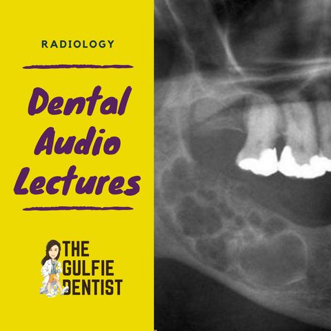
19 AGO 2020 · TYPES OF RADIOGRAPHY
IOPA
Intra – oral periapical radiograph
Function – to determine
o Working length
o Periapical pathosis QN
o External root resorption
Commonly used size – size 2
In anterior teeth – proximal caries – IOPA is used QN
If improper horizontal angulation – OVERLAPPING PROXIMAL SURFACES
19 AGO 2020 · BISECTING ANGLE
The film is placed as close as possible to the surface.
Central ray is directed perpendicular to the imaginary bisector.
Advantage – shorter exposure time, more patient acceptance cos we don’t need a beam aligner ring.
Disadvatage – image distortion/ magnification is possible. Angulation problems maybe, coz beam aligner right.
PARALELLING BEAM
It is easy to place using a film holder.
Central ray is perpendicular to both recptor & long axis of the tooth.
To maintain the parallelism between the object & the film, - INCREASE TARGET TO OBJECT DISTANCE *
Advantage – more accurate & Simplicity (no extra calculations required)
Disadvantages – patient discomfort.
19 AGO 2020 · BITE WING
Function – proximal caries** best detected by bitwing – post (whereas anterior proximal caries best – IOPA) ***
Vertical angulation for a bitewing radiograph is: 10 degree downward.
Determination of 2° caries
Crest of interdental bone
Incorrect horizontal angulation will lead to overlapping of proximal surface of teeth
19 AGO 2020 · OCCLUSAL
Commonly used in right angled technique
Function to determine
o Mesiodense
o Mandibular tori
o Sialolithiasis in Submandibular duct
o (other sialolithiasis best detected by – radiography)
It is the only radiograph can be done intraorally in TRISMUS cases QN
19 AGO 2020 · OPG
ORTHOPANTAMOGRAM
Most commonly used for dental implants QN ( BEST is CBCT )
ANATOMICAL LANDMARKS
o Mental foramen – radiolucency at the apex of premolar
o Zygoma – J shaped radio opacity at 1st or 2nd max molar
o Sinus lining
o Mandibular canal
o Vertebrae – cervical
o Styloid process
o Ear lobe
o Hyoid
ERRORS IN OPG
HEAD placed ———— ———— APPEARANCE
a) Anterior to focal trough ** Blur and narrow
b) Posterior to focal trough Blur and Magnified
19 AGO 2020 · HEAD tilted ————————— APPEARANCES
a) Downward smiling appearance to OPG Rx – tilt chin up***
b) Upward reverse curve / flat curve in OPG Rx – tilt chin DOWN***
TONGUE –
a) OBSCURES THE APICES OF MAX TEETH – SO MUST BE PLACED ON THE PALATE TO AVOID THIS !**
These are lectures of The Gulfie Dentist Online Coaching
Informazioni
| Autore | Dr.Mayakha Mariam |
| Organizzazione | Dr.Mayakha Mariam |
| Categorie | Corsi |
| Sito | - |
| thegulfiedentist@gmail.com |
Copyright 2024 - Spreaker Inc. an iHeartMedia Company
